Research Pursuits of the Macro-Molecular and Radiation Chemistry Group


The research activities of the Macro-Molecular & Radiation Chemistry Group are focused on the synthesis and functionalization of nanomaterials for the applications in areas of catalysis, optics, bio-sensing and imaging.

We develop chemical methods for synthesis of new types of inorganic nanoclusters, hybrid nano-architectures, and inorganic/polymer nanocomposites. Particular emphasis is given towards radiation-based synthesis of nanomaterials. Extensive characterizations of the physical and chemical properties of nanoparticles are carried out with microscopy and spectroscopy techniques.

Our research programme is broadly divided into following directions.


Color Centers in Diamonds: Nitrogen Vacancy Center and Beyond
The defects or impurity in the diamond crystal have attracted much attention for research around the world. Diamond strongly modifies the electronic structure of the impurity atoms which give rise to novel properties. Due to a very large band gap, diamond can accommodate a variety of optically active defects known as “color centers”. One of most studied such color center is nitrogen-vacancy center (NV center). It is formed by the incorporation of a single nitrogen atom in the diamond lattice located adjacent to a vacant carbon site. Photoluminescence spectrum of an ensemble of NV centers shows the NV0 zero phonon line (575 nm), the NV− zero phonon line (638 nm), and NV− vibrational side bands (630–800 nm).

More than 500 color centers in diamond are known, but only a few of them have been characterized properly. Only a fraction of the elements of the periodic table have been implanted into diamond. One of the topics of our group is to introduce novel color center by ion irradiation with controlled spatial distribution in diamond and study their emerging fundamental magneto-optical properties.


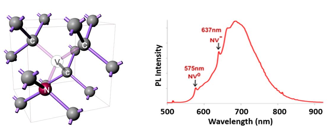
Schematic of NV center and photoluminescent spectra of nanodiamond particles containing NV centers.


Synthesis, photophysical study of graphene oxide and their interactions with biomolecular systems

Through this project we have developed improved synthetic protocols for the synthesis of graphene oxide (GO), its derivatives, composites and study their photo-physical properties, catalytic activity. We have demonstrated a facile liquid phase exfoliation method for synthesis of graphene quantum dots(GQDs) which shows excitation independent strong emission with quantum yield of 14%. As-synthesized GQDs displays delayed fluorescence and steady state anisotropy, which make the material appropriate candidate for application in sensing, bioimaging of cells and organism. We also study their interactions with biomolecular systems and their effect on the conformation of the biomolecules, such as DNA, flavo-enzymes using optical spectroscopy, isothermal titration calorimetry (ITC) etc.


Luminescent Semiconductor Nanoparticles Emitting in Second Near-Infrared Window
Fluorescent probes emitting in the second near-infrared window (NIR-II, 1000–1350nm) enable in vivo imaging of biological process with deep tissue penetration, high spatial and temporal resolution.

However, most of the reported NIR-II nanoprobes, such as PbS, PbSe, PbTe, InP and CdHgTe uses highly toxic compounds which limits their applications in vivo . Through this project we develop NIR fluorescent QDs with low toxicity for bioimaging applications.

Following is our recent results on Ag2S QDs emitting in NIR-II window.

Synthesis route of Ag2S nanoparticles.

(a) Absorbance and (b) PL spectra of the silver sulfide nanoparticles synthesized at 30OC, 50OC and 70OC.


Synthesis, Characterization and Photo-physical Properties Studies of ZnSe QDs and Their Interaction with Biomolecules
Zinc Selenide(ZnSe) QDs is an attractive semiconducting material with large band gap and attracted considerable attention owing to its wide applications in laser diodes, green-blue light-emitting diodes and solar cells.

We synthesize water soluble ZnSe QDs capped with various ligands, such as proteins, dendrimers etc.

We study the role of conduction band potential on electron transfer from QDs to metal ions or other ligands.

We also investigate the interactions of ZnSe QDs with biomolecules and nanomaterials, such as folic acid, graphene QDs etc. using optical spectroscopy, isothermal titration calorimetry to name a few.


Metal Naoclusters
The metal nanoparticle with size comparable to the de Broglie wavelength of electrons opens an energy band gap and gain molecule-like properties: discrete energy levels, absorption bands and appearance of fluorescence. These ultrasmall sized nanoparticles are often called nanoclusters (NCs). Metal(M) nanoclusters (Cu, Ag, Au etc.) attracted attention of researchers during the last decade due to their photoluminescence, good photostability, high emission rates, large Stokes’s shift, extremely high surface-to-volume ratio and low toxicity. Due to the ultrasmall size, the ligands for MNCs preparation influences on their optical properties. Currently, we are studying influence of surface ligands on optical properties of MNCs and the interaction of the MNCs nanoparticles in an assembly.


Nanoparticles Emitting in NIR-II (1000-1700 nm) Region
In the past few decades, the field of photoluminescence based biomedical imaging have developed rapidly and enabled early diagnosis, treatment, and evaluation of diseases progression. However, fluorescence imaging in the visible range and the first near-infrared (NIR-I) window (650−900 nm) faces a critical challenge in in-vivo imaging due to its limited tissue penetration depth. Imaging in NIR-II(1000−1700 nm) window can achieve unprecedented penetration depth and resolution and improve signal-to-noise ratios significantly compared to imaging in NIR-I region due to less scattering by tissue and negligible autofluorescence in this region. However, the lack of biocompatible highly fluorescent, non-toxic, easily functionalisable probes in this NIR-II window has prevented the use of this highly sensitive spectral range for in-vivo imaging. We are developing biocompatible NIR-II imaging probes with narrow-band emissions, enhanced photostability, high quantum yield and fine tune their size and composition-dependent optical properties.

Home